IJCRR - 7(14), July, 2015
Pages: 57-60
Print Article
Download XML Download PDF
ABLATION OF SULCUS SPERMATICUS FOR BIRTH CONTROL SURGERY IN MUGGER CROCODILES(CROCODYLUS PALUSTRIS)
Author: Justin William B., G. Dhananjaya Rao, M. Bharathidasan, R. Thirumurugan, S. Simon, R. Jayaprakash, Ravi Sundar George
Category: Healthcare
Abstract:Objectives: Mugger crocodiles breed twice in a year under captivity resulting in over population; requiring birth control measures. As the testis and vas deferens are intra-abdominal, the efficacy of surgical ablation of sulcus spermaticus that transport the sperm during copulation, on the dorsal surface of the phallus was assessed.
Methods: Twelve male crocodiles weighing between 140 to 230 kg were immobilized and anaesthetized with a combination of xylazine-ketamine at the dose rate of 1.5 and 20 mg/kg body weight respectively. Under aseptic precautions the phallus was retracted through cloacal slit as the animal was positioned on dorsal recumbency. The mucous membrane guarding the groove and floor were resected to ablate the sulcus spermaticus and the mucous membrane on either side was sutured. After 6 to 8 months randomly 6 crocodiles were immobilized and the phallus was examined, which revealed no signs of regeneration of the groove. Conclusion: The study revealed that ablation of sulcus spermaticus could be followed as a birth control surgery in male mugger crocodiles without affecting the welfare and breeding behavior.
Keywords: Birth control surgery, Crocodiles, Male, Mugger, Phallus, Sulcus spermaticus, Surgical ablation
Full Text:
INTRODUCTION
The mugger crocodile is a medium-sized crocodile which is restricted to the Indian sub-continent and has a broad snout, belonging to the genus Crocodyus(Silva da and Lenin, 2010). The mugger crocodiles are a hole-nesting species and the egg-laying takes place during annual dry season. Females become sexually matured when they reach approximately 1.8 to 2.0 meters and they lay 25 to 30 eggs in each clutch and the incubation period is 55 to 75 days (Whitaker, and Whitaker, 1979). In captivity they are known to lay two clutches in a single year (Whitaker and Whitaker, 1984). Often the population goes beyond the desired level which can be managed in captivity. Captive breeding and rearing programs in India have met with success. Excess numbers of captive-bred animals now reside in captivity, due to lack of suitable release sites in recent years. In reorganization of zoo rules 1992 (Wild Life-Protection-Act 1972) it is recommended to safeguard against uncontrolled growth in the population of prolifically breeding animals and every zoo shall implement appropriate population control measures like separation of sexes, sterilization, vasectomy, tubectomy and implanting of pallets etc. . The common procedures adopted in male to prevent deposition of sperm in female genitalia are either occlusion or resection of vas deferens and deviation or hiding the penis from being introduced. In crocodiles the testis and vas deferens are intra-abdominal and the vas deferens opens at the base of the penis. The measures adopted to control breeding in these species are segregation of male and female which make them more aggressive and stealing and destroying the eggs which are of welfare concern. Literature and careful anatomical study of male crocodile penis – the phallus, revealed that the presence of a sulcus spermaticus; located on the dorsal aspect of penis and is similar to urethra in depositing the sperm in female genitalia during mating (Ziegler and Olbort, 2007).The study was carried out to assess the efficacy of the surgical removal or ablation of the groove; sulcus spermaticus under anaesthesia as a birth control measure in male mugger crocodiles without affecting their breeding behaviour.
MATERIALS AND METHODS
The study was conducted on twelve male mugger crocodiles belonging to Arignar Anna Zoological Park, whose weights ranged from 140 to 230 kg, through a project on Health cover to captive Wild Animals (Prolifically Breeding Wild animals) in reorganization of zoo rules 1992, Government of India. The crocodiles were physically restrained with snares and chemically immobilized and anaesthetized with a combination of xylazine at the rate of 1.5 mg/kg body weight and ketamine at the rate of 20 mg/kg body weight administered intramuscularly (Shiju et al., 2012).
Exteriorization of phallus and examination
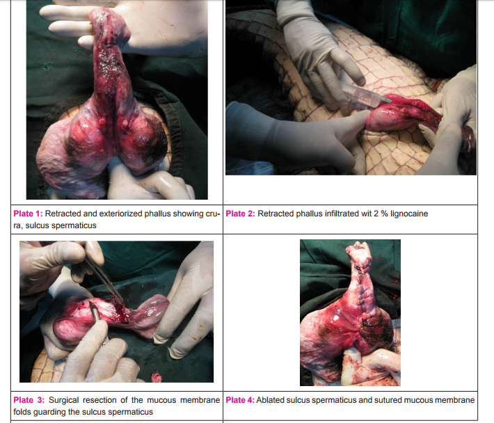
Under dorsal recumbency the ventral abdomen around the cloacal slit was cleaned and a gloved hand was introduced into the cloacal slit and the anterior aspect was explored for the presence of the phallus. The phallus in male is differentiated from female rudimentary phallus or clitoris in relation to age and length of the crocodile. The phallus was gently grasped at the crura, retracted back and exteriorized through the cloacal slit(Figure 1) to expose the shaft of the penis. The sulcus spermaticus was examined and the organ and cloaca was cleaned with one per cent povidone iodine solution.
Ablation of sulcus spermaticus
The mucus membrane folds on either side of the grove were infiltrated with 2 per cent lignocaine (Figure 2) and resected over the entire length upto glans penis using an electrocautery. The mucus membrane in the grove was excised to expose the underlying collagenous shaft with Bard-parker blade No. 10 (Figure 3). Haemorrhage was controlled and the mucous membrane on either side was opposed with polyglycolic acid suture material No. 1 without involving crura of penis (Figure 4). Povidone iodine ointment was applied on the wound edges and the phallus was repositioned in-situ.
Post-operative care
The operated mugger crocodiles were housed separately and observed for 7 days. Enrofloxacin was administered intramuscularly at the rate of 5 mg/kg body weight for three days post-operatively. The sulcus spermaticus ablated male crocodiles were left in their regular tank after 7 days along with their mates.
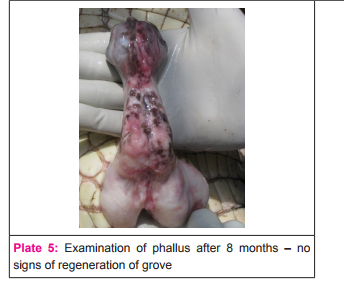
Examination of phallus after 6 months
After an interval between 6 and 8 months, 6 crocodiles were immobilized and the phallus was exteriorized and examined for the regeneration of the groove (Figure-5) under xylazineketamine immobilization. Further they were allowed to mate where only the sulcus spermaticus ablated males were retained in the tank along with the female.
RESULTS
Xylazine at the rate of 1.5 mg/kg and ketamine at the rate of 20 mg/kg when administered intramuscularly induced chemical immobilization and anaesthesia for a period of 45±3.26 minutes and the animals recovered in 2.45±0.91 hours.
Phallus anatomy
The phallus was hidden inside the longish cloacal slit. The retracted phallus was an unpaired laterally compressed organ located on the ventral wall of cloaca. The phallus could be divided into three regions; proximally a pair of crura located dorsal to the ischia, a fused penile shaft and distal glans. The penile shaft was curved and a V-shaped groove; the sulcus spermaticus, formed by a pair of mucous membrane folds that acted as wall on either sides of the groove. The groove extended the full length of the penile shaft and glans on the dorsal side when the phallus was in-situ. The groove was deep around one centimetre in adult mugger and it became shallow as it reached the glans penis. The glans penis was lappet-shaped and the tip was irregular with mucous membrane folds to which the sulcus spermaticus extended in the form of small branches (Figure-1,2). The mucous membrane cover over the shaft and gland penis was loosely adherent to the underlying tissue.
Ablation of sulcus spermaticus
Resection and excision of the mucous membrane folds on either side of the sulcus spermaticus resulted in total ablation of the groove. After opposing the mucous membrane the conical shape of the phallus was maintained. Bleeding was effectively controlled by the use of thermocautery. During the post-operative period the animals did not show any signs of discomfort.
Examination of phallus after 6 months
Examination of phallus in 4 crocodiles after 6 to 8 months of surgery revealed no signs of regeneration of the groove (Figure-5). Mating resulted in unfertilized without hatchlings following natural incubation.
DISCUSSION
Phallus anatomy and ablation of sulcus spermaticus
In mugger crocodiles a pair of testes (testis), are situated in the abdominal cavity on either sides of the spinal column. Vas deferens arising from testis, passes from the dorsal side of the cloaca to the ventral side, not accompanied by the ureter and opens into the blind sac which forms the basal continuation of the deep groove on the dorsal side of the phallus. As the testis and vas deferens are intra-abdominal, vasectomy could be performed only through laparotomy which was not attempted because it could be a major surgery with expected post-operative complications and the position of anterior abdominal vein. In certain lizards the vas deferens and ureter unite and form a short canal which opens beneath or upon a small papilla(Das and Purkayastha, 2012). Hence attempt was not made to close the blind sac as it could interfere with excretion from the urinary system and if the anatomical nature was similar to lizards in muggers. The copulatory organ in crocodiles is unpaired and called a phallus. It is formed of cavernous tissue and is erected when engorged with blood. The penis consists of two parallel bands of cancellous tissue, the corpora cavernosa (corpora cavernosa penis) at the ventral wall of the cloaca. Between them lies a groove, the sulcus spermaticus, which transports sperm from the vasa deferens. There is a glans (glans penis) of cavernous tissue at the extremity of the phallus. When engorged with blood, the corpora cavernosa enlarge and shape the sulcus spermaticus into a duct through which sperm flows and protrude the glans from the cloacal vent enabling its insertion into the cloaca of the female (Kelly, 2013). Amputation of penis was performed in a Nile crocodile for recurrent paraphimosis and bleeding (Lankester and Hernandez, 2005). When the animal was on dorsal recumbency; retraction of crura and exteriorization of phallus gave good visualization of the groove on the ventral aspect of the animal making it convenient for the surgery. The haemorrhage was controlled by electrocautery and suturing.
Examination of phallus after 6 months
Occasional cases of tail regeneration have been observed in crocodilians (Alibardi, 2010). In the present study regeneration could not be observed upto 8 months. Allowing the crocodiles with ablated sulcus did not produced fertilized eggs as no hatchlings could be located.
CONCLUSION
Ablation of sulcus spermaticus on the phallus of mugger crocodiles resulted prevented deposition of sperm into the female genitalia during mating without affecting the breeding behaviour and upto 8 months follow-up; the sulcus spermaticus did not regenerated.
ACKNOWLEDGEMENTS
The authors acknowledge the help rendered by the Director of Clinics, TANUVAS, Dean, Madras Veterinary College and Director of Arignar Anna Zoological Park, Chennai for the support and financial help for the purchase of drugs and surgical disposable. The authors acknowledge the immense help received from the scholars whose articles are cited and included in references of this manuscript. The authors are also grateful to authors / editors / publishers of all those articles, journals and books from where the literature for this article has been reviewed and discussed.
Competing Interests The authors declare that they have no competing interests.
References:
1. Alibardi, L., 2010. Regeneration of reptiles and its position among vertebrates. In. Morphological and cellular aspects of tail and limb regeneration in Lizards. A model system with implications for tissue regeneration in mammals. Springer, p1-49.
2. Das, M and Purkayastha, J., 2012. Insight into hemipenial morphology of five species of Hemidactylus Oken, 1817 (Reptilia: Gekkonidae) of Guwahati, Assam, India, Hamadryad. 36(1): 32 – 37.
3. Kelly, D.A., 2013. Penile anatomy and hypotheses of erectile function in the American Alligator (Alligator mississippiensis): Muscular eversion and elastic retraction. The Anat. Rec., 296: 488-494.
4. Lankester, F. and Hernandez, DSJ., 2005. Paraphimosis and amputation in a Nile crocodile (Crocodylus niloticus).J. Zoo Wildl. Med., 36(4):698-701.
5. Shiju, S, William, BJ., Jayaprakash, R.; Kumar, RS. and Thejomoorthy, P, 2012. Evaluation of xylazine-ketamine and xylazine - acepromazine -ketamine for surgical intervention in mugger crocodiles (Crocodylus palustris). Indian J. Field Vet. 7(4): 41- 44.
6. Silva da, A.and Lenin, J., 2010. Mugger crocodile Crocodylus palustris. In: Crocodiles. Manolis, S.C. and C. Stevenson (Eds), 3rd Edn., Staus Survey and Conservation Action Plan, India. P94-98.
7. Whitaker, R. and Whitaker, Z., 1979. Preliminary crocodile survey – Sri Lanka, J. Bombay Nat. Hist. Soc. 76 (1) 66-85. 8. Whitaker, R. and Whitaker, Z., 1984. Reproductive biology of mugger. J. Bombay Nat. Hist. Soc. 81(2) 119-127.
9. Wild Life (Protection) Act 1972, Recognition of Zoo Rules, 1992. Government of India.
10. Ziegler, T., and Olbort, S., 2007. Genital structures and sex identification in Crocodiles. Crocodile Specialist Group Newsletter, 26(3): 16-17.
|
 IJCRR
IJCRR 


 This work is licensed under a Creative Commons Attribution-NonCommercial 4.0 International License


