IJCRR - 5(20), October, 2013
Pages: 121-124
Date of Publication: 02-Nov-2013
Print Article
Download XML Download PDF
UNUSUAL LOCATION OF CORACOBRACHIALIS MUSCLE AND COURSE OF MUSCULO CUTANEOUS NERVE - A CASE REPORT
Author: Kalpana T., Udaya Kumar P., Murali Krishna S., Rajesh V., Chandra Mohan M., Naveen Kumar B.
Category: Healthcare
Abstract:Musculocutaneous nerve typically pierces the coracobrachialis muscle. During gross anatomy dissections of the upper extremities, for freshman undergraduates, in the department of Anatomy, Mamata Medical College, Andhra Pradesh, the coracobrachialis muscle was found to be innervated by a nerve branch arising from the lateral cord of the brachial plexus. The musculocutaneous nerve was found to course downwards medial to coracobrchialis and biceps brachii muscles and then pierce the bceps brachii instead of coracobrachialis muscle. The coracobrachialis was found in deeper plane, posterior to short head of biceps brachii rather than medial to it. No other abnormality was observed in the branching pattern of the brachial plexus on both sides. Knowledge of the anatomical variations of the location of coracobrachialis and peripheral nervous system is important to clinicians and surgeons in interpreting unusual clinical presentations.
Keywords: Coracobrachialis, Biceps brachii, Musculocutaenous nerve, Brachial plexus.
Full Text:
INTRODUCTION
Coracobrachialis takes origin from the coracoid process, together with the tendon of the short head of biceps and inserts on to the medial border of the shaft of the humerus. This muscle forms an inconspicuous rounded ridge on the upper medial side of the arm. Usually the muscle is perforated by the musculocutaneous nerve. Biceps and brachialis muscles are related laterally to this muscle whereas pectoralis major, brachial vessels and median nerve are related anteriorly1.
The musculocutaneous nerve arises from the lateral cord (C5–7) of brachial plexus. It pierces coracobrachialis and descends laterally between biceps and brachialis to the lateral side of the arm. Just below the elbow it pierces the deep fascia lateral to the tendon of biceps, and continues as the lateral cutaneous nerve of the forearm. It supplies coracobrachialis, both heads of biceps and most of brachialis muscle1.
CASE REPORT
During routine dissection labs for freshman undergraduate students of Mamata Medical College, Andhra Pradesh, a variation of abnormal location of coracobachialis muscle and unusual course of musculo cutaneous nerve in relation the muscle is observed in 60 yr old male cadaver.
Morphological variations of coracobrahialis muscle are common but the variation in the location of muscle was not reported so far. In this present case coracobrachialis muscle was found originating from the medial border of coracoid process of scapula along with the tendon of short head of biceps. Then muscle coursed unusually deep and posterior to short head of biceps brachii muscle rather than descending medial to it. Insertion of the muscle was observed on the anterior surface of mid shaft of humerus instead of medial surface. A nerve branch from the lateral cord was found to innervate the muscle near its origin rather than from musculocutaneous nerve.
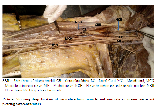
Musulo cutaneous nerve was observed arising from lateral cord of brachial plexus as usual. Interestingly the nerve did not pierce the coracobrachialis, instead continued downwards medial to it and then passed between the biceps and brachialis muscles after giving nerve branches to both (Picture No 1). Nerve supply to coraco brachialis was derived from lateral cord of brachial plexus rather than from musculo cutaneous nerve. The muscular braches to biceps and brachialis were originated from musculocutaneous nerve. Finally the musculocutaneous nerve continued as lateral cutaneous nerve of forearm).
DISCUSSION
This case report presents the abnormal location of coracobrachialis muscle and unusual continuation of musculo cutaneous nerve without piercing the coracobrachialis muscle in right upper limb of 60 yr old Indian male cadaver.
Jakubowicz2, Kopuz C3, Mehmet4, Nakatani5, Lee6, Mostafa7 and Sargon8 observed an accessory head of coracobrachialis but loactation of coracobrachialis as deep and posterior to biceps short head and its insertion on the anterior surface of shaft of humerus has not been reported previously.
Absence of musculocutaneous has been reported by many authors. Prasad rao et al9, Pacholczak R et al10, Guerry Guttenberg RA11, Natakani et al12, Jamuna et al13, Uzel AP14 reported unilateral absence of musculocutaneous nerve and Ihunwo et al15 observed the same bilaterally. In the present case the nerve was observed to be arising from the lateral cord as usual.
Authors Guerry Guttenberg11, Gumusalan16, Himabindu17, Jamuna13, Chitra18, Nayak19and Natakani20 observed that the musculo cutaneous nerve did not pierce the coracobrachialis in its course, rather found it passing downwards and medial to the muscle. They observed that the nerve either ended up by joining the median nerve or continued as lateral cutaneous nerve of forearm. In the present study also musculo cutaneous nerve did not pierce the muscle instead continued downwards medial to the biceps muscle then entered it and finally continued as lateral cutaneous nerve of forearm. No communicating branches were observed between musculo cutaneous nerve and median nerve.
CONCLUSION
Upper extremity is a frequent site of injury and various surgical and invasive procedures. Coracobrachialis muscle has been suggested for use in coverage in infraclavicular defects of postmastectomy reconstructive patients21 and also in free vascularized muscle transfer for treatment of longstanding facial paralysis22. So knowledge of such anatomical variations in muscles and nerves of upper limb is of utmost importance to clinicians.
Competing Interests
The authors declare that we have no competing interests
Ethical committee clearance
As the study included only human cadavers, ethical committee clearance was not taken into consideration. Authors will take the responsibility of any further allegations regarding ethical clearance that arise from the study.
ACKNOWLEDGEMENTS
I thank my colleagues and supporting staff for their valuable suggestions and support. I extend my gratitude to all the scholars / authors / editors / publishers whose articles, journals are reviewed, cited and included in the references of this manuscript.
References:
- Standring S, Borley NR, Collins P, Crossman AR, Gatzoulis MA, Healy JC, et al., Gray’s anatomy. 40th Ed. Londaon; Churchill Livingstoe, 2008.
- Jakubowicz M, Ratajczak W., Variation in morphology of the biceps brachii and coracobrachialis muscles associated with abnormal course of blood vessels and nerves. Folia Morphol (Warsz). 2000; 58(4):255-8.
- Kopuz C, Içten N, Yildirim M., A rare accessory coracobrachialis muscle: a review of the literature. Surg Radiol Anat. 2003 Feb; 24(6):406-10.
- Taylor GI, Cichowitz A, Ang SG, Seneviratne S, Ashton M., Comparative anatomical study of the gracilis and coracobrachialis muscles: implications for facial reanimation. Plast Reconstr Surg. 2003 Jul; 112(1):20-30.Mehmet Mutlu Catli, Umut Ozsoy, Yasemin Kaya, Arzu Hizay, Fatos Belgin Yildirim, and Levent Sarikcioglu., Four-headed biceps brachii, three-headed coracobrachialis muscles associated with arterial and nervous anomalies in the upper limb, Anat Cell Biol. 2012 June; 45(2): 136–139.
- Nakatani T, Tanaka S, Mizukami S., Bilateral four-headed biceps brachii muscles: the median nerve and brachial artery passing through a tunnel formed by a muscle slip from the accessory head. Clin Anat. 1998;11: 209–212.
- Lee SE, Jung C, Ahn KY, Nam KI., Bilateral asymmetric supernumerary heads of biceps brachii. Anat Cell Biol. 2011;44:238–240.
- Mostafa M El-Naggar., A study on the morphology of the coracobrachialis muscle and its relation with the musculocutaneous nerve, Folia Morphol; Vol,60, No.3, pp.217-224
- Sargon MF, Tuncali D, Celik HH., An unusual origin for the accessory head of biceps brachii muscle. Clin Anat. 1996;9:160–162
- Prasada Rao PVV, Chaudahary SC., Absence of musculocutaneous nerve: two case reports. Clin Anat 2001: 14:31-5
- Pacholczak R, Klimek - Piotrowska W, Walocha J A., Absence of the musculocutaneous nerve associated with a supernumerary head of biceps brachii: a case report. Surg Radiol Anat. 2011 Aug; 33(6):551-4.
- Guerri - Guttenberg R A, Ingolotti M., Classifying musculocutaneous nerve variations., Clin Anat. 2009 Sep;22(6):671-83.
- Nakatni T, Mizukami S, Tanaka S., Absence of the musculocutaneous nerve with innervation of coracobrachialis, the biceps brachii, the brachialis and lateral border of the forearm by the branches from the lateral cord of the brachial plexus. J Anat 1997b; 191:459-60
- Jamuna M, Amudha G., A cadaveric Study on the anatomic variations of musculocutaneous nerve in the infra clavicular part of brachial plexus, Journal of clinical and diagnostic research (2011) Nov (suppl-1), Vol 5(6): 1144-1147
- Uzel AP, Bulla A, Steinmann G, LaurentJoye M, Caix P., Absence of the musculocutaneous nerve and its distribution from median nerve: About two cases and literature review, Morphologie. 2011 Dec; 95(311):146-50.
- Ihunwo AO, Osinde SP, Mukhtar AU., Disstribution of median nerve to the muscles of the anterior compartment of rhe arm. Cent Afr J Med 1997;43:359-60
- Gümü?alan Y, Yazar F, Ozan H, Variant innervation of the coracobrachialis muscle and unusual course of the musculocutaneous nerve in man, Kaibogaku Zasshi. 1998 Jun; 73(3):269-72.
- Himabindu.A, Narsinga Rao. B., short musculocutaneous nerve and unusual innervations of median nerve., Int J of BAMS, 2012 may, Vol 2 (2): 166-169
- Chitra R. Multiple bilateral neuroanatomical variations of the nerves of the arm. Neuroanatomy 2007; 6:43-5
- Nayak S, Samuel VP, Somayaji N: Concurrent variations of the median, the musculo cutaneous nerve and the biceps brachii muscle. Neuroanatomy 2006; 5:30-2
- Nakatani T, Mizukami S, Tanaka S., Three cases of the musculocutaneous nerve not perforating the coracobrachialis muscle. Kaibogaku Zasshi. 1997 Jun;72(3):191-4.
- P C Hobar, R J Rohrich, T J Mickel., The Coracobrachialis muscle flap for coverage of exposed axillary vessels; a salvage procedure. Plastic and amp. recon. surg 06/1990; 85(5): 801-4
|
 IJCRR
IJCRR 


 This work is licensed under a Creative Commons Attribution-NonCommercial 4.0 International License


