IJCRR - 5(20), October, 2013
Pages: 109-115
Date of Publication: 02-Nov-2013
Print Article
Download XML Download PDF
MORPHOMETRIC STUDY OF HUMAN ADULT CADAVERIC KIDNEYS-RESEARCH ARTICLE
Author: Sivanageswara Rao Sundara Setty, Raja Sekhar Katikireddi
Category: Healthcare
Abstract:Aim: The main aim of the study was to perform morphometric analysis of right and left kidney specimens and compare findings with the previous reports. Materials and Methods: A total of 50 human adult cadaveric kidneys (25 right and 25 left) were studied. Morphometric features like length, breadth, width and weight were measured. Hilum and lobulation of kidneys were observed. Results: The weight of right kidneys varied from 57 to 190 gms with an average of 103.04 gms. The weight of left kidney ranged from 60 to 220 gms with an average of 114.48 gms. The length of right kidney varied between 8 and 14 cms with an average of 10.92 cms. The length of left kidney varied between 9.5 and 14.5 cms with an average of 11.32 cms. The breadth of the right kidney ranged between 5 and 8 cms with an average of 6.2 cms. The breadth of left kidneys was in the range of 5 to 9 cms with an average of 6.62 cms. The width of right kidney varied between 2 and 5 cms with an average of 3.34 cms. The width of left kidney varied between 2 and 5 cms with an average of 3.54 cms. Lobulation is present in 8% of right kidney and 16% of left kidney specimens. Variations in hilar structures were found in 8% of right kidney and 24% of left kidney specimens. Conclusion: Anatomical variations of kidney rely on morphological findings. Clinically, this could play an important role in improving the knowledge of anatomists, surgeons and radiologists.
Keywords: Kidney, Hilum, Length, Breadth, Width.
Full Text:
INTRODUCTION
Kidneys are a pair of chief excretory organs that not only maintain the electrolyte and water balance but also serve as endocrine organs [1]. They are considered as retroperitoneal organs and are situated in the posterior abdominal wall beside the vertebral column and extend from T1 to L3 vertebra [2].
Each kidney is bean shaped and has a length of 11 cms, breadth of 6 cms and width of 3 cms. The left kidney is 1.5 cm longer than the right. The average weight of a kidney is 150 grams [1, 2]. Kidneys are characterized by a circular and thick superior pole and a pointed and thin inferior pole. The anterior surface is convex and posterior surface is flat [2]. The lateral border is convex, medial border is concave with a hilum that consists of renal vein, renal artery and pelvis of the ureter, anterio-posteriorly [2]. Foetal lobulation could persist in the adult life such that the renal outline appears larger than the normal [3].
In the recent period, morphometric studies have gained much research attention as they are believed to possess significant clinical importance. Most probably, variations related to renal dimensions observed in such studies are anticipated to furnish better insights on anomalies.
For instance, conditions like systemic diseases, urinary tract diseases, congenital anamolies, neoplasia, micro and macrovascular diseases were reported to significantly influence kidney sizes [4]. With the advent of imaging technology, small renal tumors were also being identified [5]. In addition, structural arrangements or alterations at the hilum also possess medical significance, as per the available case reports [5-8].
But, studies related to morphometirc determination of renal dimensions and hilum structures still appear limited and need to be strengthened with the additional findings. Therefore, the main objective of the study is to carry out morphometric study of human adult cadaveric kidneys and compare with the data reported elsewhere.
Materials and Methods
The present study was conducted in the Anatomy Department of Bhaskar Medical College, Yenkapally Village, Moinabad Mandal, Ranga Reddy district, Andhra Pradesh. A total of 25 right and 25 left human adult cadaveric kidneys were included. Initially, the shapes of kidneys were noted. Then using an electronic weighing machine, kidneys were weighed.
Length, breadth and width of kidneys were measured with the help of digital sliding calipers and noted. We studied the structures present at the hilum of the kidneys. We considered the maximum distance between the two poles of the kidneys as its length, maximum distance between two points at the same level between the medial and lateral borders as its breadth and maximum width as the width of kidneys. We also studied the lobulation of the kidneys. The data obtained was tabulated, analysed statistically and compared with the previous studies.
Results
Out of 50 kidneys studied, 25 were right kidneys and 25 were left kidneys. All the 50 kidney specimens were bean shaped. Among the 25 right kidneys, weight ranged from 57 to 190 gms and the average weight was found to be 103.04 gms (Table 1.) The length of right kidney varied between 8 and 14 cms with an average length of 10.92 cms (Table 1). The breadth of right kidneys was in the range of 5 to 8 cms, with an average breadth of 6.2 cms (Table 1). The width of the right kidney varied between 2 and 5 cms with an average width of 3.34 cms (Table 1).
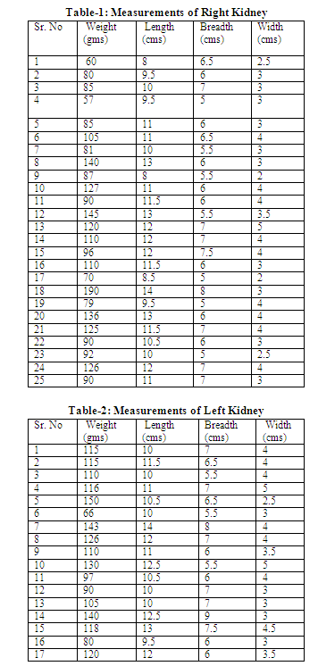
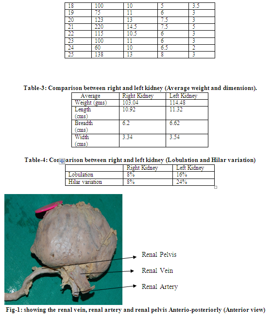
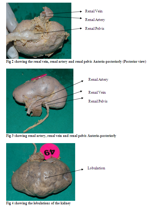
Among 25 right kidney specimens, 2 (8%) showed lobulation (Table 4). We observed hilum of the right kidney with an arrangement of renal vein, renal artery and renal pelvis anterio-posteriorly in 23 (92%) specimens. In contrast, 2 (8%) specimens showed renal artery, renal vein and renal pelvis anterio-posteriorly (Table 4).
Among the 25 left kidneys, the weight ranged from 60 to 220 grams and the average weight was found to be 114.48 gms (Table 2). The length of left kidneys varied between 9.5 and 14.5 cms with an average length of 11.32 cms (Table 2). The breadth of left kidneys was in the range of 5 to 9 cms with an average breadth of 6.62 cms (Table 2). The width of left kidneys varied between 2 and 5 cms with an average width of 3.54 cms (Table 2). Among 25 left kidney specimens, 4(16%) showed lobulation (Table 4). We observed the hilum of left kidney with an arrangement of renal vein, renal artery and renal pelvis anterio-posteriorly in 19 (76%) of specimens. The remaining 6(24%) left kidney specimens showed renal artery, renal vein and renal pelvis anterio-posteriorly at the hilum (Table 4). The normal and variational renal hilar structures are represented in figures 1-3 and lobulation of kidneys are represented in the figure 4.
Discussion
Kidneys are the important organs to maintain the homeostatic function of the body and act as endocrine organs [1]. The present study was done to explore morphological variations of right and left kidneys and describe their significance. We noted several variations in the kidney morphology.
In the present study, all the 50 (100%) kidneys were bean shaped as mentioned in the standard text books of Anatomy [1,2]. The average weight of right kidneys was 103.04 gms and the average weight of the left kidneys was 114.48 gms. This is not coinciding with the earlier studies that described the average weight to be 108.7 +/- 22.6 g and 111.8 +/- 23.3 g for right and left kidneys, respectively [9]. This is also not in agreement with some studies where the average weight was taken into consideration commonly for both kidneys [1, 10]. This could indicate that our present study showed a variation in the weight of kidneys when compared with the earlier findings.
Next, kidney size is considered as an important indication for many clinical signs and hence it is worth studying. Previous studies showed that aging leads to a progressive decrease in kidney size, especially after middle age [11,12]. Recently, a significant correlation between kidney size and kidney function was observed in patients with chronic kidney disease (CKD) [13].
In the present study, right kidney size measurements revealed an average length of 10.92 cms, average breadth of 6.2 cms and the average width of 3.34cms. These are closer to earlier findings [4, 14] but varied with some other [10]. Similarly, left kidney size measurements revealed
an average length of 11.32 cms, breadth of 6.62 cms and the width of 3.54 cms which are closer to the previous findings [1] but differed from some other studies [10]. So, this indicated variations in the renal dimensions and could generate considerable medical interest.
It could be possible that the renal dimensions might also vary among population of different geographical origin. However, as not much data is available, renal variations need further exploration. Our further emphasis was on lobulation and renal hilum. We observed lobulation in 8% of right kidney specimens and 16% of left kidney specimens. Normally, the foetal kidneys are subdivided into lobules which disappear during infancy as the nephrons increase and grow [15]. Patil and his associates reported a rare congenital condition of kidney where bilateral lobulation and malrotation were observed in association with the open hilar structure of kidney [16]. The lobulation observed in the present study although had no associations with any other structural variations or defects, it might highlight certain clinical significance.
We observed variations in the renal hilar structures both in the right and left kidneys, but more at the left kidney (24%). Generally, renal hilum variant patterns were reported to be frequent on the left kidney [5]. This could be attributed to the developmental defects of the renal veins.
From the embryological view point, right renal vein develops from one channel whereas
left renal vein develops from several anastomotic channels. Any abnormality during these channel developments could alter the arrangement of renal hilar structures with regard to the renal vein [5]. So, our findings related to renal hilar structural variation is supported by the previous studies [5-8].
Overall, we observed that weight, dimensions, hilar structural variations and lobulations of left kidney were larger than the right kidney (Table 3). This could indicate that left kidney is more susceptible to anatomical variations than the right kidney.
Conclusion
In conclusion, renal dimensions and hilar structural arrangements could possess significant clinical value. It is necessary to distinguish a pathological kidney from a normal sized healthy kidney. Determination of renal anatomical variants should be greatly encouraged to strengthen the current literature and improve the knowledge needed for surgical and radiological interventions.
Acknowledgements
We are thankful to Dr. K. V. Vijayasaradhi (Prof.), Dr. Mahopatra (Prof.), Dr. Hima Bindu (Assoc. Prof), Dr. Mohd. Abid Ali (Asst. Prof), Dr. S. Parimala (Asst. Prof) and Dr. B. Sirisha (Asst. Prof), Bhaskar Medical College, for their kind cooperation and coordination.
References:
- Standring S: Gray’s Anatomy: The Anatomical Basis of the Clinical Practice, 39th edition. Edinburg: Elsevier Churchill Livingstone, 2006; 1269-84.
- Datta AK. Essentials of Human Anatomy (Thorax and Abdomen) part I, 7th ed., Calcutta: Current books international, 2006; 301-320.
- Datta AK. Essentials of Human Embryology part, 5th ed., Calcutta: Current books international, 2005; 220-224.
- Raza, M, Hameed, A and Khan, I. Ultrasonographic Assessment of Renal Size and its Correlation with Body Mass Index in Adults Without known Renal Disease. J Ayub Med Coll Abbottabad 2011;23(3):64-68
- Trivedi, S.; Athavale, S. & Kotgiriwar, S.Normal and Variant Anatomy of Renal HilarStructures and its Clinical Significance. Int. J. Morphol, 2011;29(4):1379-1383.
- Das S, Paul S,. Variation of renal hilar structures: A cadaveric case.Eur J Anat 2006;10 (1): 41-43
- Naveen Kumar, Ashwini P, Aithal, Anitha Guru, Nayak Satheesha B. Bilateral vascular variations at the renal hilum: a case report. Case Rep Vasc Med 2012;2012:968506.
- Verma P, Arora AK, Sharma P, Mahajan A. Variations in branching pattern of renal artery and arrangement of hilar structures in the left kidney: clinical correlations, a case report. Ital J Anat Embryol. 2012;117(2):118-22.
- Sahni D, Jit I, Sodhi L. Weight and measurements of kidneys in northwest Indian adults. Am J Hum Biol13 (6):726-32.
- Khatun, H, Sultana Z, Islam, N, Kibria, Chy, T. Morphological Study of the Kidney in Relation to Age. Bangladesh Journal of Anatomy 2009;7(1):19-21
- Mileti? D, Fuckar Z, Susti? A, Mozetic V, Stimac D, Zauhar G. Sonographic measurement of absolute and relative renal length in adults J Clin Ultrasound, 1998; 26(4):185-9.
- Akpinar IN, Altun E, Avcu S, Tüney D, Ekinci G, Biren T. Sonographic measurement of kidney size in geriatric patients. J Clin Ultrasound, 2003;31(6):315-8.
- Jovanovi? D, Gasic B, Pavlovic S, Naumovic R.Correlation of kidney size with kidney function and anthropometric parameters in healthy subjects and patients with chronic kidney diseases.Renal Failure 2013;35(6): 896-900
- Sampaio F J, Mandarim-de-Lacerda CA.Morphometry of the kidney. Applied study in urology and imaging. J Urol (Paris) 1989;95 (2):77-80.
- Keith. L. Moore. Clinically Oriented Embryology, 8th ed. Keith Lean Moore, Trivedi V. N. Persaud,
- Patil ST, Meshram MM, Kasote AP. Bilateral malrotation and lobulation of kidney with altered hilar anatomy: a rare congenital variation. Surg Radiol Anat 2011;33(10):941-4.
|
 IJCRR
IJCRR 


 This work is licensed under a Creative Commons Attribution-NonCommercial 4.0 International License


