IJCRR - 5(21), November, 2013
Pages: 50-55
Date of Publication: 21-Nov-2013
Print Article
Download XML Download PDF
STUDY OF TRANSVERSE AND SAGITTAL DIAMETERS OF LUMBAR PEDICLES IN RELATION TO TRANS-PEDICULAR SCREW FIXATION USING MRI IN RAJASTHAN POPULATION
Author: Meghna Bhaumik, Neelam Bapna, Uday Bhaumik, K. Prabhakaran
Category: Healthcare
Abstract:Introduction: The technique of inter-pedicular screw fixation for the purpose of spinal fixation has gained enormous importance in the recent years. The choice of screw for the procedure is determined by the minimum (transverse) diameter of the pedicle .Morphometric data on the diameters of the pedicles are therefore useful in preoperative planning and in designing pedicle screws.Most of the previous study of the morphometry of pedicle is based on white population using x-rays and CT scans. Therefore the present study was conducted in the Western region of India in state of Rajasthan to measure the morphometry of pedicles in relation to trans-pedicular screw fixations as well as to distinguish differences in the morphometry of pedicles in men and women of different age groups using MRI technique. Aims and Objectives: To evaluate the sexual differences in transverse and Sagittal diameters of lumbar vertebral pedicle. Also to determine the difference of diameter in the pedicles of different age groups and finally to determine the morphometry of pedicles in relation to trans- pedicular screw fixation in the region of Rajasthan. Materials And Methods: MRI scan of 1000 patients (both males and females) between the age group of 20 to 60 years, with the history of low back pain reporting to the outpatient department of Neurosurgery, Orthopedics, Rehabilitation and inpatients admitted in various wards for complaints of disc protrusion, disc extrusion, spondolysthesis (not associated with gross vertebral body collapse) of S.M.S. hospitals, Jaipur formed the material for the current study. Results: There is a cranio\?caudal increase of transverse and sagittal diameters from L1 toL5 vertebrae. Dimensions of male population are significantly higher than female population with respect transverse diameter and sagittal diameter. With the above parameters the size of the screw for transpedicular screw fixation can be derived.
Keywords: lumbar vertebra, lumbar pedicle, trans-pedicular screw fixation
Full Text:
INTRODUCTION
The technique of inter-pedicular screw fixation for the purpose of spinal fixation has gained enormous importance in the recent years.
Stefee et al1 and Zindric2 described the screw fixation procedure as the method of choice for stabilization of the lumbo-sacral spine. Many types of pedicle screw systems have been developed; basically they all entail the insertion of screws through the pedicle from the posterior aspect into the vertebral bodies. The success of the technique depends upon the ability of the screws to obtain and maintain balance within the vertebral body2 .This is determined among other factors by the accuracy of choice of screw, size of the pedicle and the quality of the bone of the pedicle.
The choice of screw for the procedure is determined by the minimum (horizontal) diameter of the pedicle3,4 .Morphometric data on the diameters of the pedicles are therefore useful in preoperative planning and in designing pedicle screws.
Most of the previous study of the morphometry of pedicle is based on white population5-10
Almost all the previous workers have reported the data on morphometry of the pedicle based on a common pool of vertebrae where male and female vertebrae were pooled together5,6,2,7. However, recently Amonoo Kuofi10 has reported statistically significant sex differences in pedicle morphometry.
Thus according to Kragman3, as the racial variations in skeleton are well known the morphometry of pedicle may vary population to population, even within the same population, the anatomical variations have been reported in the shape, size and angulations of the pedicles by Weinstein et al4.
C.T. scans or M.R.I. sections represent a new approach to vertebral morphometry allowing certain measurements to be measured for the first time.
Morphometric studies of the pedicles have demonstrated variations in pedicle shape, size and angulations. Carefully taken routine radiographs, CT scans and MRI scans can help in determine about the diameter, length and angulations of the pedicle. This is critically important, particularly in placing screws in rotated vertebrae and deformed spines like in patient with scoliosis or spondylolisthesis.
Anatomic variations may prevent placing a screw in a particular pedicle or necessitate using a smaller screw than originally planned.
Risks of neural damage can be minimized by thorough knowledge of the anatomy of vertebral column as well as practicing on anatomical specimens. Therefore the present study was conducted in the Western region of India in state of Rajasthan to measure the morphometry of pedicles in relation to trans-pedicular screw fixations as well as to distinguish differences in the morphometry of pedicles in men and women of different age groups.
AIMS AND OBJECTIVES
- To evaluate the sexual differences in transverse and
- Sagittal diameters of lumbar vertebral pedicle.
- To determine the difference of diameter in the pedicles of different age groups.
- To determine the morphometry of pedicles in relation to trans- pedicular screw fixation in the region of Rajasthan.
MATERIALS AND METHODS
MRI scan of 1000 patients (both males and females) between the age group of 20 to 60 years, with the history of low back pain reporting to the outpatient department of Neurosurgery, Orthopedics, Rehabilitation and inpatients admitted in various wards for complaints of disc protrusion, disc extrusion, spondolysthesis (not associated with gross vertebral body collapse) of S.M.S. hospitals, Jaipur formed the material for the current study. Only Patients who were natives of Rajasthan State (born and brought up in Rajasthan) were included in the study.
Whereas Patients suffering from congenital spinal deformities like Achondroplasia, Tethered cord syndrome, spinal dysraphism, spilt cord malformation etc. and Patients affected with spinal tuberculosis, spinal meningitis and even Patients coming with spinal trauma along with bony injuries, were excluded from the study. Also Patients below 20 years of
age and above 60 years of age who were not natives of Rajasthan were excluded from the present study. The study design is mainly of descriptive type.
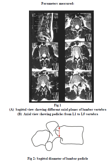
The measurement of the lumbar pedicles of one thousand patients was recorded with the magnetic resonance imaging (M.R.I. scanning).
The measurements of angles in MRI scans were measured using diacom software.
Student’s t test was used for statistical analysis.
DISCUSSION
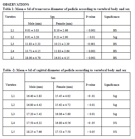
Transverse diameter of pedicle – The transverse diameter of pedicles are showing a steady increase from first to fifth lumbar vertebrae. The value of transverse diameter of pedicle is 9.01 mm at L1 vertebra and 18.00 mm at vertebra L5, while in females the minimum value is 8.10 mm at L1 vertebra and the maximum value is 16.01 mm at L5 vertebra. P value is highly significant for L1, L3, L4 and L5 vertebrae (P<0.001), between male and female population and significant changes are noted at L2 vertebra in both sexes. With advancing age this diameter also follows a steady increase in dimensions.
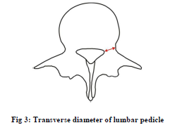
Sagittal diameter of pedicle – This parameter is showing increase in dimensions of pedicles in cranio -caudal directions with the minimum values of 16.40 mm in males at L1 vertebra to the maximum value of 18.25 mm at L5 vertebra, while in females the minimum value of sagittal diameter is 15.45 mm at L1 vertebra and the maximum value of sagittal diameter is 17.53 mm at L5 vertebra. Significant changes (P<0.01) are recorded between male and female populations, while non significant changes are noted at L4 and L5 vertebrae in both sexes. Sagittal diameter also undergoes linear increase in dimensions as the age progresses.
The studies conducted by H.S. Amonoo Kuofi10, Mitra et al11 on horizontal and vertical diameters of pedicles stated, that the values of sagittal diameter followed an increasing trend from above downwards (from vertebra L1 to L5), the highest value for sagittal diameter was found at L5 vertebra and the lowest value was for vertebra L1. The values of sagittal diameters of pedicles in males were 17.1 at L1 vertebra and in females it was 16.2 mm10 and the sagittal diameter was 18.6 mm in males and in females it was 17.65 mm at vertebra L510.
In present study we have also noted, values regarding sagittal diameters of pedicles similar to Amonoo Kuofi10. In present study the sagittal diameter of pedicles in males is 16.40 mm and in females it is 15.45 mm for vertebra LI. At vertebra L5 the sagittal diameters of pedicles in males is 18.25 mm and in females it is 17.53 mm. In present study the values of sagittal diameters are lower in females than male subjects.
CONCLUSION
From the present study following conclusions are derived –
- There is a cranio–caudal increase of transverse and Sagittal diameters from L1 toL5 vertebrae.
- Dimensions of male population are significantly higher than female population with respect transverse diameter and Sagittal diameter.
- With the above parameters the size of the screw for transpedicular screw fixation can be derived.
The large sample size of the study reiterates and establishes the observations, that there are significant differences in pedicular morphology in Indian population, when compared with Western counterparts. Although the trends are similar with few exceptions but the, values are consistently lower for various Morphometric Measurements in Indian population. The difference in values may be on account of ethnic related morphologic differences as Indians have noticeably smaller build than their Western counterparts. Ethnicity with its diverse cultural, dietary and genetic factors and not just the body size or height, has the potential to influence the overall pedicle morphometry.
Thus, this study strengthens the case for customizing the existing range of spine pedicle implants according to ethnic characteristics or developing ethnic specific implants. Further, this study indicates that computer software aided pedicular morphometery data are quite reliable and comparable to other published studies with other methods, but has advantages like being much simpler, more user friendly, unbiased and patient specific. Therefore, the need for preoperative computer aided M.R.I/C.T. base morphometrics
References:
- Steffee AD, Sitkowski DJ, Topham LS. Total vertebral body and pedicle arthroplasty. Clin Orthop Relat Res. 1986; Feb;(203):203-8
- Zindrick MR. The role of transpedicular fixation systems for stabilization of the lumbar spine. Orthop Clin North Am. 1991; Apr;22(2):333-44.
- Krag MH, Beynnon BD, Pope MH, Frymoyer JW, Haugh LD, Weaver DL. An internal fixator for posterior application to short segments of the thoracic, lumbar, or lumbosacral spine. Design and testing. Clin Orthop Relat Res. 1986; Feb;(203):75-98
- Weinsten JN, Rydevik BL, Rauschning W. Anatomic and Technical considerations of pedicle screw fixation. Clinical Orthopaedics and related research 1986; 248:34-46.
- Saillant G. Anatomical study of the vertebral pedicles. Surgical application. Rev Chir Orthop Reparatrice Appar Mot. 1976 Mar;62(2):151-60.
- Roy Camiller R, Saillant G, Lapresle P, Mazel C. A secret in spine surgery. The pedicle, 51st meeting of the American Academy of Orthopaedic Surgeons. Atlanta, Georgia, 1984.
- Berry JL, Moran JM, Berg WS, Steffee AD. A morphometric study of human lumbar and selected thoracic vertebrae. Spine. 1987; May;12(4):362-7
- Scoles PV, Linton AE, Latmimer B, Levy ME, Diglovanni BF. Vertebral body and posterior element morphology: The normal spine in middle life. Spine 1988;13:1082-1086.
- Olsewski JM, Simmon EH, Kallen FC, Mendel FC, Severin CM, Berens DL. Morphometry of lumbar spine, Anatomical perspectives related to transpedicular screw fixation. Journal of bone and joint Surgery 1990;72A: 541-549.
- Amonoo-Kuofi HS. Maximum and minimum lumbar interpedicular distances in normal adult Nigerians. J Anat. 1982; Sep;135(Pt 2):225-33.
- Mitra SR, Datir SP, Jadhav SO. Morphometric study of the lumbar pedicle in the Indian population as related to pedicular screw fixation. Spine. 2002; Mar 1;27(5):453-9.
|
 IJCRR
IJCRR 


 This work is licensed under a Creative Commons Attribution-NonCommercial 4.0 International License


